Frostbite is the damage to tissues from freezing ;The blood vessels contract and cause loss of oxygen to the affected body parts.And results formation of ice crystals within cells, rupturing the cells and leading to cell death. It most commonly affects areas that are further away from the body core and have less blood flow. These include your feet, hands, nose, and ears.
There are three degrees of cold injury: frostnip, superficial frostbite, and deep frostbite.
The affected skin may be slightly flushed. The skin changes to white or grayish yellow as the frostbite develops. Pain is sometimes felt early but subsides later. Often there is NO pain; the part being frostbitten simply feels intensely cold and numb.
In superficial frostbite, there will be an area that looks white or grayish and the surface skin will feel hard but the underlying tissue will be soft. With deeper involvement, large blisters appear on the surface, as well as in underlying tissue, and the affected area is hard, cold and insensitive. Destruction of the entire thickness of the skin will necessitate skin grafting and will constitute a medical emergency, because gangrene may result from loss of blood supply to the injured part.
In the most of neonatal resuscitations, if adequate ventilation is achieved, no need for chest compressions . However, in certain cases of advanced asphyxia and myocardial depression or severe pulmonary dysfunction in which adequate ventilation cannot be readily achieved, chest compressions are necessary to support the circulation during more extensive resuscitation.
Thus, the indication for chest compressions in the newly born differs significantly from that in older children and adults. The mechanics of the thoracic cage and the physical forces of the circulation of blood also differ, especially in preterm infants. The predominance of pulmonary dysfunction, necessitates a relatively lower ratio of compressions to ventilations. The 3:1 ratio of compressions to ventilations is performed with 90 compressions and 30 interposed breaths per minute (or one cycle of 4 events every 2 seconds).
The preferred method for chest compressions is the two-thumb-encircling-hands method , which provides firm support for the back and generates higher systemic arterial pressure and better coronary perfusion pressure than the two-finger method.
The patient may complain of eye pain or a foreign body sensation after being poked in the eye with a finger or twig. The patient may have abraded the cornea inserting or removing contact lenses. Removal of a corneal foreign body produces some corneal abrasion, but corneal abrasion can even occur without identifiable trauma. There is often excessive tearing and photophobia. Often the patient cannot open his eye for the exam. Abrasions are occasionally visible on sidelighting the cornea. Conjunctival inflammation can range from nothing to severe conjunctivitis with accompanying iritis.
What to do:
* Instill topical anesthetic drops (to permit exam). * Perform a complete eye exam (visual acuity, funduscopy, anterior chamber bright light, conjunctival sacs for foreign body). * Perform the fluorescein exam by wetting a paper strip impregnated with dry orange fluorescein dye and touching this strip into the tear pool inside the lower conjunctival sac. After the patient blinks, darken the room and examine the patient's eye under cobalt blue or ultraviolet light (the red-free light on the ophthalmoscope does not work). Areas of denuded or devitalized corneal epithelium will fluoresce green. * If a foreign body is present, remove it and irrigate the eye. * If iritis is present (evidenced by photophobia, an irregular pupil or meiosis, and a limbic blush in addition to conjunctival injection) consult the ophthalmologic followup physician about starting the patient on topical mydriatics and steroids (e.g., cyclopentolate or homatropine and prednisolone). * Instill antibiotic ointment (e.g., erythromycin, tobramycin) in the lower sac. A small, superficial, non-painful abrasion may be left uncovered. # For large, deep, and painful abrasions, patch the eye with enough pressure to keep the lid closed by folding one eyepatch double to rest against the lid, covering it with a second unfolded eyepatch, and taping both tightly with several strips of 1" tape running from the cheek to mid forehead. # Prescribe analgesics (e.g., oxycocone, ibuprofen, naproxen), and give the first dose. # Warn the patient the pain will return when the local anesthetic wears off. # Make an appointment for ophthalmologic followup to reevaluate the abrasion the next day.
What not to do:
* Do not be stingy with pain medication. Patching alone will not eliminate the pain. * Do not give patient any topical anesthetic for continued instillation. * Do not patch a patient with a bacterial conjunctivitis or ulcer. * Do not tape an eye patch up and down or across the nose.
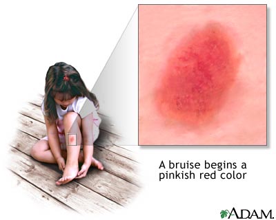
1. Red to blue: about 1 to 2 days old 2. Blue to purple: about 3 to 5 days old 3. Green: about 6 to 7 days old 4. Yellow to brown: about 8 to 10 days old 5. Resolved: at least 13 to 28 days old 6. It is likely safest to describe bruises as either: "new" (red, purple, or blue) or
"old" (green, yellow, or brown)
Note: The presence of bruises that have various ages may signify multiple episodes of injury caused by ongoing physical abuse.