The anterior approach is an approach to the front of the hip joint as opposed to a lateral (side) approach to the hip or posterior (back) approach. It is a true anterior approach to the hip and should not be confused with the Harding approach which is often referred to as an anterior approach.
Rabu, 03 Juni 2015
Selasa, 02 Juni 2015
Between Tinea capitis and Psoriasis of the scalp

- a.Androgenic hair loss
- b.Psoriasis of the scalp
- c.Seborrheic dermatitis
- d.Tinea capitis
- e.Carbuncle
The answer is d.


A mild but widespread infection by Tinea capitis (Scalp ringworm). Inflamed areas are seen on the front, behind the ear and on the back of the neck.Black dots are from broken hair
Psoriasisis a hereditary disorder characterized by scaling patches and plaques appearing in specific areas of the body, such as the scalp, elbows, lumbosacral region, and knees. The lesions are “salmon pink” with a silver-colored scale that on removal produces blood (Auspitz sign). The Koebner phenomenon (with trauma, the lesion jumps to a new location) is also elicited in patients with psoriasis.Seborrheic dermatitisis a common chronic dermatosis occurring in areas with active sebaceous glands (face, scalp, and body folds) and may occur either in infancy or in people over the age of 20. The eczematous plaques of seborrheic dermatitis are yellowish red and are often greasy with a sticky crust. Androgenic hair loss is a progressive hereditary bitemporal, frontal, or vertex balding that may begin any time after puberty. A carbuncle is a deep infectious collection of interconnecting abscesses (furuncles) arising from several hair follicles.
Uvular Necrosis after Endoscopy

The patient felt well after the procedure and was discharged home. He noted a mild sore throat, starting 24 hours after the procedure. When it persisted, he presented for evaluation.
Physical examination revealed necrosis of the distal uvula. No specific therapy was given, and acetaminophen was recommended for discomfort. The patient reported that the tip of the uvula spontaneously sloughed off the next day, and the discomfort resolved completely. He has had no further solid-food dysphagia.
Uvular necrosis is a rare event that can occur after upper endoscopy or direct laryngoscopy. The symptoms are generally mild, and the recovery is usually complete. The mechanism of injury is thought to be impingement of the uvula by the instrument against the hard palate or posterior pharynx, leading to ischemia. Uvular injury has also been reported as a result of aggressive oropharyngeal suctioning.
Senin, 01 Juni 2015
Senin, 25 Mei 2015
Causes, Symptoms and sings of aortic valvular insufficiency
A 74-year-old man presents with exertional dyspnea and generalized weakness. On examination, you discover a high-pitched, blowing diastolic murmur and a wide pulse pressure with bounding pulses. The most likely diagnosis is?
- A) aortic stenosis
- B) aortic insufficiency
- C) mitral stenosis
- D) mitral insufficiency
- E) coarctation of the aorta
Answer and Discussion
The answer is B.Causes of aortic regurgitation :

Symptoms and sings of aortic valvular insufficiency :
The symptoms of aortic valvular insufficiency are the same in older persons as they are in younger ones. Usually, the main symptoms are related to heart failure, with exertional dyspnea and weakness being common symptoms. In some elderly patients, symptoms of dyspnea and palpitations may be more common at rest than with exertion. Nocturnal angina pectoris, often accompanied by flushing, diaphoresis, and palpitations, may occur; this is thought to be related to the slowing of the heart rate and the drop of arterial diastolic pressure. The classic findings of a high-pitched, blowing diastolic murmur and a wide pulse pressure with an abruptly rising and collapsing pulse should make the diagnosis of aortic valvular insufficiency easily recognized in elderly patients.
Photo description of Pterygium of the eye


With corneal involvement, even if arrested surgically, a pterygium can affect vision by warping the surface of the cornea and inducing astigmatism, and/or by actually growing over in front of the pupil and obstructing the entering light.
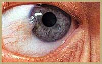 |
| Triangular, thin, transparent conjunctival fold in the palpebral fissure. Its head is yellow, avascular and pints toward the corneal center. The body of the pterygium extends to the semilunar fold(this image for a 32-year-old man). |
Treatment :
Pterygia seem to occur more frequently in people who spent much time outside, and is especially common in the southern lattitudes "Some consider pterygium as an occupational disease" So protecting the eyes from sun, dust and wind and use of lubricating drops is the standard treatment.If the pterygium is still troublesome it can be removed surgically under local anaesthetic ,with high rate of recurrence. Once the pterygium is removed, the bare sclera is covered with conjunctiva
Kamis, 21 Mei 2015
Photos and information about Blue Nevus
- Blue Nevus is a group of melanocytic lesions which all appear blue in colour due to the optical effects of light reflecting off melanin deep in the dermis.

- The blue nevus is a benign, usually solitary lesion, represents a localized proliferation of dermal melanocytes. It presents as a dark blue to black, moderately firm, rounded, sharply defined nodular tumour composed of spindle-shaped melanocytes with slender cytoplasmic processes, occurring often in association with melanin-laden macrophages in a sclerotic dermis.
- A blue naevus (nevus) is a rather unusual but non-cancerous mole.
- The blue nevus is also called the blue skin mole, or Jadassohn-Tièche nevus with 2 clinically recognized variants : the common blue nevus and the cellular blue nevus.
 - Cellular blue nevus is larger, especially on buttocks and can degenerate into malignant melanoma.
- Clinically The blue nevus appears as a dark-blue or blue-black smooth nevus formed by melanin-heavily pigmented spindle cells in the middle and lower two-thirds of the dermis. Also, the blue nevus appears as a slate blue or bluish black, sharply circumscribed, flat or slightly elevated nodule, occurring on any area of the body.
- A biopsy should be performed on any changing pigmented lesion. For a solitary lesion, simple excision is usually curative. Rare cases of persistent blue nevi, manifesting as satellite lesions around the original excision site, have been reported. These must be distinguished from malignant blue nevus, and reexcision is recommended.


- The blue nevus is a benign, usually solitary lesion, represents a localized proliferation of dermal melanocytes. It presents as a dark blue to black, moderately firm, rounded, sharply defined nodular tumour composed of spindle-shaped melanocytes with slender cytoplasmic processes, occurring often in association with melanin-laden macrophages in a sclerotic dermis.
- A blue naevus (nevus) is a rather unusual but non-cancerous mole.
- The blue nevus is also called the blue skin mole, or Jadassohn-Tièche nevus with 2 clinically recognized variants : the common blue nevus and the cellular blue nevus.

- Clinically The blue nevus appears as a dark-blue or blue-black smooth nevus formed by melanin-heavily pigmented spindle cells in the middle and lower two-thirds of the dermis. Also, the blue nevus appears as a slate blue or bluish black, sharply circumscribed, flat or slightly elevated nodule, occurring on any area of the body.
- A biopsy should be performed on any changing pigmented lesion. For a solitary lesion, simple excision is usually curative. Rare cases of persistent blue nevi, manifesting as satellite lesions around the original excision site, have been reported. These must be distinguished from malignant blue nevus, and reexcision is recommended.

Common blue nevus
Langganan:
Postingan (Atom)